The fold of the left vena cava, ligament of the left vena cava, or vestigial fold of Marshall, is a triangular fold of the serous pericardium that lies...
1 KB (132 words) - 15:34, 17 May 2024

The valve of the inferior vena cava (Eustachian valve) is a venous valve that lies at the junction of the inferior vena cava and right atrium. In prenatal...
5 KB (683 words) - 16:01, 26 February 2024

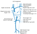
The inferior vena cava is a large vein that carries the deoxygenated blood from the lower and middle body into the right atrium of the heart. It is formed...
10 KB (1,079 words) - 16:37, 4 May 2024
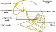
superior vena cava and the ascending aorta. Bachmann's bundle is, during normal sinus rhythm, the preferential path for electrical activation of the left atrium...
4 KB (474 words) - 14:21, 18 May 2024
Atrium (heart) (redirect from Left atrium)
responsible for venous drainage; it derives from the embryonic left superior vena cava. During embryogenesis at about two weeks, a primitive atrium begins...
23 KB (2,481 words) - 15:15, 15 May 2024

Chordae tendineae (redirect from Tendon of Todaro)
Eustachian valve of the inferior vena cava and the valve of the coronary sinus. Along with the opening of the coronary sinus and the septal cusp of the tricuspid...
8 KB (788 words) - 07:26, 3 July 2024

Pericardial sinus (category Wikipedia articles incorporating text from the 20th edition of Gray's Anatomy (1918))
pulmonary trunk , and anterior to the superior vena cava. This sinus is clinically important because passing one end of clamp through the sinus, and the other...
4 KB (369 words) - 07:36, 28 January 2023
Crista terminalis (redirect from Crista terminalis of His)
terminalis of His) is a vertical ridge on the: 56 posterolateral inner surface of the adult right atrium extending between the superior vena cava, and the...
4 KB (448 words) - 21:53, 8 April 2024
the sinus of the vena cava, or sinus venarum cavarum) is the portion of the right atrium in the adult human heart where the inner surface of the right...
2 KB (207 words) - 17:56, 22 October 2023

the superior/inferior vena cava and pulmonary veins enter the heart. The root of the great vessels and the associated reflections of the serous pericardium...
13 KB (1,465 words) - 14:49, 17 October 2023
primary pacemaker of the heart. It is a region of cardiac muscle on the wall of the upper right atrium near to the superior vena cava entrance. The cells...
11 KB (1,462 words) - 20:56, 2 July 2024

Ventricle (heart) (redirect from Left ventricle)
pumping of blood throughout the body and lungs is much greater than the pressure generated by the atria to fill the ventricles. Further, the left ventricle...
20 KB (2,368 words) - 21:09, 27 April 2024

tissue or organ of the body. The coronary arteries wrap around the entire heart and both lungs . The two main branches are the left coronary artery and...
11 KB (1,265 words) - 16:34, 28 March 2024
Heart valve (redirect from Left cusp of aortic valve)
pulmonary artery. The heart also has a coronary sinus valve and an inferior vena cava valve, not discussed here. The heart valves and the chambers are lined...
22 KB (2,655 words) - 01:39, 24 June 2024
Coronary circulation (redirect from Left posterior aortic sinus)
and other components of the heart. Two coronary arteries originate from the left side of the heart at the beginning (root) left ventricle. There are three...
16 KB (1,977 words) - 21:49, 28 November 2023

wide, and 1 mm thick, located directly below and to the side of the superior vena cava. These cells can produce an electrical impulse known as a cardiac...
24 KB (2,806 words) - 17:50, 24 April 2024
Fossa ovalis (heart) (redirect from Limbus of fossa ovalis)
of the right atrium. If the aneurysm stretches too far, it can narrow the opening of the inferior vena cava. This type of aneurysm can be a result of...
7 KB (907 words) - 15:30, 18 January 2024
formed by as semicircular fold of the lining membrane of the right atrium. It is situated at the base of the inferior vena cava.[citation needed] The valve...
4 KB (373 words) - 19:58, 18 June 2024

primary pacemaker) positioned on the wall of the right atrium, near the entrance of the superior vena cava. Other pacemaker cells are found in the atrioventricular...
42 KB (5,101 words) - 08:00, 30 June 2024
Mitral valve (redirect from Left atrioventricular valve)
or left atrioventricular valve, is one of the four heart valves. It has two cusps or flaps and lies between the left atrium and the left ventricle of the...
20 KB (2,337 words) - 22:32, 8 April 2024

Cardiac conduction system (redirect from Conducting system of the heart)
the left and right bundle branches of His to the respective Purkinje fibers for each side of the heart, as well as to the endocardium at the apex of the...
16 KB (1,840 words) - 02:31, 30 January 2024
heart of humans and most other animals, located between the left ventricle and the aorta. It is one of the four valves of the heart and one of the two...
12 KB (1,504 words) - 01:26, 24 June 2024
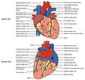
Heart (redirect from Left side of the heart)
the right and left atrium continuously. The superior vena cava drains blood from above the diaphragm and empties into the upper back part of the right atrium...
143 KB (16,888 words) - 17:57, 17 June 2024

serous fluid secreted by the serous layer of the pericardium into the pericardial cavity. The pericardium consists of two layers, an outer fibrous layer and...
5 KB (530 words) - 13:07, 4 March 2023
the anterior-inferior end of the interatrial septum, to the ventricles of the heart. The bundle of His branches into the left and the right bundle branches...
7 KB (772 words) - 00:43, 21 December 2023

Bundle branches (redirect from Left anterior fascicle)
The left bundle branch further divides into the left anterior fascicle and the left posterior fascicle. These structures lead to a network of thin filaments...
4 KB (506 words) - 08:44, 11 December 2023
Purkinje fibers (redirect from Fibers of Purkinje)
portion of the cardiac cycle, the Purkinje fibers carry the contraction impulse from both the left and right bundle branch to the myocardium of the ventricles...
8 KB (839 words) - 02:21, 1 April 2024

the coronary sinus near its left extremity; it is continuous above with the ligament of the left vena cava (vestigial fold of Marshall), and the two structures...
1 KB (130 words) - 14:56, 8 May 2024
artery, with the remainder originating from the left circumflex artery. This is associated with the dominance of the coronary artery circulation. In right-dominant...
11 KB (1,218 words) - 17:51, 24 April 2024
Intervenous tubercle (redirect from Tubercle of Lower)
superior vena cava toward the atrioventricular opening. This article incorporates text in the public domain from page 531 of the 20th edition of Gray's...
1,000 bytes (83 words) - 12:34, 25 January 2020