
Ribbon diagrams, also known as Richardson diagrams, are 3D schematic representations of protein structure and are one of the most common methods of protein...
12 KB (1,073 words) - 19:39, 30 May 2024
States Ribbon diagram (or Richardson diagram), 3D schematic representation of protein structure Ribbon (film), a 2017 Indian film Ribbon, Kentucky Ribbon knot...
2 KB (265 words) - 14:43, 16 July 2024

developing the Richardson diagram, or ribbon diagram, a method of representing the 3D structure of proteins. Ribbon diagrams have become a standard representation...
33 KB (3,244 words) - 15:18, 30 September 2024

Ribbon diagram of a monomer of human MAO-A, with FAD and clorgiline bound, oriented as if attached to the outer membrane of a mitochondrion. From PDB:...
37 KB (4,019 words) - 14:53, 25 September 2024
Ribbon diagram of a mouse antibody against cholera that binds a carbohydrate antigen...
102 KB (11,386 words) - 22:54, 1 October 2024

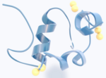
deathstalker's powerful venom contains the 36-amino acid peptide chlorotoxin (ribbon diagram shown). This blocks small-conductance chloride channels, immobilizing...
91 KB (9,238 words) - 06:21, 22 September 2024

Streptavidin Monomeric streptavidin (ribbon diagram) with bound biotin (spheres); PDB: 1STP Identifiers Organism Streptomyces avidinii Symbol ? UniProt...
20 KB (2,487 words) - 20:35, 13 August 2023

Thaumatin family Ribbon diagram of thaumatin I. From PDB: 1RQW. Identifiers Symbol Thaumatin Pfam PF00314 InterPro IPR001938 SMART SM00205 PROSITE PDOC00286...
11 KB (1,219 words) - 12:56, 3 September 2024

region 3 (CDR3), which forms an extended loop (coloured orange in the ribbon diagram above) covering the lipophilic site that normally binds to a light chain...
35 KB (3,898 words) - 14:53, 18 May 2024

Alkaline phosphatase Ribbon diagram (rainbow-color, N-terminus = blue, C-terminus = red) of the dimeric structure of bacterial alkaline phosphatase. Identifiers...
55 KB (6,147 words) - 11:12, 12 September 2024

achieve a target structure, researchers first developed a two-dimensional diagram and utilized it to determine the constraints that allowed them to construct...
8 KB (823 words) - 10:21, 10 October 2024

Green fluorescent protein ribbon diagram...
12 KB (1,462 words) - 13:12, 21 September 2023
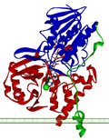
monoamine oxidase A Ribbon diagram of a monomer of human MAO-A, with FAD and clorgiline bound, oriented as if attached to the outer membrane of a mitochondrion...
38 KB (4,085 words) - 19:06, 30 September 2024

Molecule editor Polyhedral skeletal electron pair theory Quantum chemistry Ribbon diagram Styx rule (for boranes) Topology (chemistry) McMurry, John E. (1992)...
23 KB (2,314 words) - 11:20, 14 September 2024

Ribbon diagram of a protease (TEV protease) complexed with its peptide substrate in black with catalytic residues in red.(PDB: 1LVB)...
26 KB (2,799 words) - 15:02, 30 June 2024
hydrogen white, oxygen red, and nitrogen blue. On the right side is a ribbon diagram of the insulin hexamer, believed to be the stored form. A monomer unit...
121 KB (13,799 words) - 17:37, 8 October 2024
A ribbon diagram depicting the crystal structure of the GPIbα N-terminal domain including the VWF A1 and thrombin binding sites....
14 KB (1,648 words) - 22:47, 2 December 2023

A ribbon diagram of apolipoprotein E. Variants of this protein influence the risk of developing DLB....
136 KB (14,528 words) - 00:36, 9 October 2024
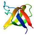
SH3 domain Ribbon diagram of the SH3 domain, alpha spectrin, from chicken (PDB accession code 1SHG), colored from blue (N-terminus) to red (C-terminus)...
8 KB (933 words) - 12:56, 10 September 2024

A ribbon diagram of the MCP of Pseudoalteromonas virus PM2, with the two jelly roll folds colored in red and blue...
25 KB (2,893 words) - 04:22, 4 July 2023

The human androgen receptor bound to testosterone The protein is shown as a ribbon diagram in red, green, and blue, with the steroid shown in white....
209 KB (21,568 words) - 16:45, 2 October 2024

Trp repressor protein Ribbon diagram of the trpR protein Identifiers Symbol Trp_repressor Pfam PF01371 Pfam clan CL0123 InterPro IPR000831 SCOP2 2wrp /...
7 KB (759 words) - 00:04, 15 February 2023
A ribbon diagram of the MCP of Pseudoalteromonas virus PM2, with the two jelly roll folds colored in red and blue...
30 KB (3,641 words) - 00:53, 17 August 2024

Ribbon diagram of human carbonic anhydrase II, with zinc atom visible in the center...
40 KB (4,383 words) - 22:18, 6 August 2024
Molecular graphics (section Ribbon diagrams)
Ribbon diagrams are schematic representations of protein structure and are one of the most common methods of protein depiction used today. The ribbon...
28 KB (2,002 words) - 07:14, 9 September 2024

EPSP Synthase (3-phosphoshikimate 1-carboxyvinyltransferase) Ribbon diagram of EPSP synthase liganded with shikimate (spheres). Identifiers EC no. 2.5...
12 KB (1,366 words) - 05:06, 23 September 2024

protease inhibitor nirmatrelvir bound to the viral 3CLpro protease enzyme. Ribbon diagram of the protein with the drug shown as sticks. The catalytic residues...
25 KB (2,009 words) - 15:17, 10 October 2024

Ribbon diagram of human carbonic anhydrase II, with zinc atom visible in the center...
143 KB (16,228 words) - 07:01, 9 October 2024

Ribbon diagram of Shiga toxin (Stx) from S. dysenteriae. From PDB: 1R4Q....
19 KB (2,311 words) - 12:39, 5 October 2024

Botulinum toxin A Ribbon diagram of tertiary structure of BotA (P0DPI1). PDB entry 3BTA. Clinical data Trade names Botox, Myobloc, Jeuveau, others Other...
116 KB (11,608 words) - 02:54, 12 October 2024