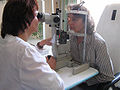
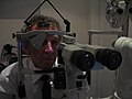
In ophthalmology and optometry, a slit lamp is an instrument consisting of a high-intensity light source that can be focused to shine a thin sheet of...
20 KB (2,550 words) - 22:39, 15 July 2024

Eye examination (section Slit-lamp)
Fluorescein staining before slit lamp examination may reveal corneal abrasions or herpes simplex infection. The binocular slit-lamp examination provides stereoscopic...
18 KB (2,186 words) - 22:04, 11 October 2024
of the lateral chamber angle. The method involves a narrow slit of light from a slit lamp being projected onto the peripheral cornea at an angle of 60⁰...
11 KB (1,311 words) - 08:52, 5 June 2024

Wilson's disease (section Slit-lamp examination)
rings) may be visible in the cornea of the eyes, either directly or on slit lamp examination as deposits of copper form a ring around the cornea. This...
42 KB (4,960 words) - 01:34, 16 September 2024

with a slit lamp. A fundus lens or macular lens is used to aim beam of light on the macula. It is best performed in a dilated eye, projecting a slit beam...
6 KB (505 words) - 19:24, 18 May 2024

fingernails. About 25% of cases occur at work. Diagnosis is often by slit lamp examination after fluorescein dye has been applied. More significant injuries...
18 KB (2,175 words) - 02:40, 7 November 2023

angle), using a goniolens (also known as a gonioscope) together with a slit lamp or operating microscope. Its use is important in diagnosing and monitoring...
7 KB (854 words) - 11:05, 24 May 2024

or whether treatment can be accomplished without additional resources. Slit lamp examination is invaluable in diagnosis but initial assessment can be performed...
14 KB (1,505 words) - 21:49, 25 July 2024
who need to lie flat, or patients unable to co-operate with a sitting slit lamp examination, that yields clinical results comparable to the Goldmann....
20 KB (2,296 words) - 09:25, 25 May 2024

by professor Arthur Korn in 1902, and in Allvar Gullstrand's original slit lamp (1911) which is used for ophthalmology to allow physicians to view the...
5 KB (560 words) - 15:09, 17 October 2024

neurofibromatosis is done via the following means: Radiograph MRI or CT scan EEG Slit-lamp examination Genetic testing Histology Conditions similar to NF include:...
25 KB (2,366 words) - 03:39, 13 October 2024
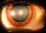
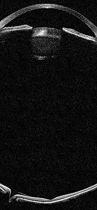
Magnified view of a cataract seen on examination with a slit lamp...
151 KB (15,178 words) - 01:45, 3 October 2024

needed] Genes include: The diagnosis of Fuchs dystrophy is often made with slit lamp biomicroscopy. With direct illumination, the clinician can visualize guttae...
16 KB (1,738 words) - 19:53, 23 August 2024

conjunctiva, manifested by irritation and redness. Examination using a slit lamp (biomicroscope) may improve diagnostic accuracy. Examination of the palpebral...
38 KB (3,942 words) - 08:05, 9 October 2024
determine intraocular pressure Refraction assessment Retina examination Slit lamp examination Visual acuity Color vision test Visual field test Dry eye...
59 KB (7,250 words) - 00:57, 11 October 2024

affect vision, but are very useful in diagnosis. They are detected by slit lamp examination. Immunohistochemistry stains positive against the proteins...
4 KB (337 words) - 00:53, 11 September 2024
Slit lamp photo of posterior capsular opacification visible a few months after implantation of intraocular lens, seen on retroillumination...
64 KB (6,615 words) - 12:52, 9 October 2024

cornea in a pinpoint pattern, which can be seen with examination with a slit-lamp. Patients may present with non-specific symptoms such as red eye, tearing...
2 KB (177 words) - 01:14, 31 January 2023

Ancillary tests for abnormal structures and physiology can be made via a slit lamp test, which examines the cornea, conjunctiva, anterior chamber, and iris...
23 KB (2,171 words) - 04:46, 1 October 2024

Unilateral or bilateral subepithelial lesions of the cornea may be present. Slit lamp examination reveals multiple tiny granular deposits surrounded by a halo...
2 KB (160 words) - 12:34, 17 January 2024
observed via fundoscopy (using an ophthalmoscope to view inside the eye) or slit lamp exam. The original retinal spots identified in 1872 were attributed to...
5 KB (366 words) - 22:00, 6 September 2024
Holly. The conventional and most common TBUT measurement method is using slit lamp and sodium fluorescein. Noninvasive instruments (Oculus Keratograph 5M...
10 KB (1,167 words) - 12:51, 19 September 2024
slit lamp biomicroscopy to examine the transparent axial tissues of the eye. Traditionally, ophthalmic biomicroscopy has been completed with a slit lamp...
10 KB (1,311 words) - 07:39, 30 March 2023

pupils. A slit lamp examination will also be conducted to examine the cornea, iris, and other areas closer to the front of the eye. A slit lamp is used...
8 KB (979 words) - 00:27, 23 March 2024
will search for other characteristic findings of the disease by means of slit lamp examination of the cornea. An advanced case is usually readily apparent...
66 KB (7,263 words) - 18:13, 3 September 2024

conjunctival microvessels are typically imaged with a high-magnification slit lamp with green filters. With such high-magnification imaging systems, it is...
20 KB (1,900 words) - 15:34, 27 August 2024
Right esotropia Rx Prescription SE Spherical Equivalent SLE Slit lamp examination SLM Slit lamp microscope EP Esophoria ET Esotropia Supp. Suppression V...
13 KB (38 words) - 06:30, 8 March 2023

dark-brown, and may be complete or broken. The rings are best seen using the slit lamp under cobalt blue filter. They are named for Bruno Fleischer. Fleischer...
4 KB (301 words) - 07:14, 18 August 2024
present under slit-lamp examination. Iritis can occur, too. Keratic precipitates are another eye manifestation (detectable by a slit lamp, but are usually...
112 KB (11,757 words) - 19:47, 9 September 2024

ache and head ache may be present. Drooping of eyelid may be present. Slit lamp examination shows loss of epithelium. The site of the ulcer is studied...
13 KB (1,557 words) - 07:44, 1 September 2024