
microscopy: optical, electron, and scanning probe microscopy, along with the emerging field of X-ray microscopy.[citation needed] Optical microscopy and...
69 KB (8,315 words) - 10:01, 9 July 2024

Atomic force microscopy (AFM) or scanning force microscopy (SFM) is a very-high-resolution type of scanning probe microscopy (SPM), with demonstrated...
75 KB (9,806 words) - 17:28, 9 July 2024

Fluorescence microscope (redirect from Flourescence microscopy)
more complex fluorescence microscopy techniques like confocal microscopy and total internal reflection fluorescence microscopy while xenon lamps, and mercury...
25 KB (2,745 words) - 10:17, 9 August 2024

Scanning electron microscope (redirect from Scanning electron microscopy)
specialized instruments. An account of the early history of scanning electron microscopy has been presented by McMullan. Although Max Knoll produced a photo with...
66 KB (8,138 words) - 15:40, 28 June 2024
Microscopy and Microanalysis is a peer-reviewed scientific journal that covers original research in the fields of microscopy, imaging, and compositional...
2 KB (104 words) - 03:39, 27 April 2023

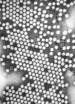
Electron microscope (redirect from Electron microscopy)
Transmission electron microscopy (TEM) where swift electrons go through a thin sample Scanning transmission electron microscopy (STEM) which is similar...
49 KB (5,605 words) - 04:15, 8 August 2024
Transmission electron microscopy (TEM) is a microscopy technique in which a beam of electrons is transmitted through a specimen to form an image. The specimen...
118 KB (15,058 words) - 21:08, 30 July 2024
Phase-contrast microscopy (PCM) is an optical microscopy technique that converts phase shifts in light passing through a transparent specimen to brightness...
12 KB (1,190 words) - 09:47, 1 June 2024

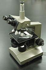
Optical microscope (redirect from Optical microscopy)
optical microscopy which do not use visible light include scanning electron microscopy and transmission electron microscopy and scanning probe microscopy and...
52 KB (5,972 words) - 03:47, 11 April 2024
interference microscopy Differential interference contrast microscopy Fluorescence interference contrast microscopy Interference reflection microscopy Phase...
446 bytes (40 words) - 21:45, 18 July 2023
Confocal microscopy, most frequently confocal laser scanning microscopy (CLSM) or laser scanning confocal microscopy (LSCM), is an optical imaging technique...
46 KB (5,298 words) - 06:21, 9 April 2024

Stimulated emission depletion (STED) microscopy is one of the techniques that make up super-resolution microscopy. It creates super-resolution images by...
33 KB (3,866 words) - 17:27, 8 June 2024

Scanning tunneling microscope (redirect from Electron tunnel microscopy)
Tunneling Spectroscopy (STS)", Scanning Probe Microscopy: Atomic Force Microscopy and Scanning Tunneling Microscopy, NanoScience and Technology, Berlin, Heidelberg:...
47 KB (7,166 words) - 23:26, 29 June 2024
Scanning probe microscopy (SPM) is a branch of microscopy that forms images of surfaces using a physical probe that scans the specimen. SPM was founded...
29 KB (3,376 words) - 14:27, 7 May 2024
Super-resolution microscopy is a series of techniques in optical microscopy that allow such images to have resolutions higher than those imposed by the...
87 KB (10,123 words) - 03:18, 29 July 2024

Cryogenic electron microscopy (cryo-EM) is a cryomicroscopy technique applied on samples cooled to cryogenic temperatures. For biological specimens, the...
23 KB (2,514 words) - 18:42, 4 August 2024
Expansion microscopy (ExM) is a sample preparation tool for biological samples that allows investigators to identify small structures by expanding them...
17 KB (2,099 words) - 09:16, 25 October 2022

Tomography (redirect from Synchroton-radiation X-ray tomographic microscopy)
methods listed above. A new technique called synchrotron X-ray tomographic microscopy (SRXTM) allows for detailed three-dimensional scanning of fossils. The...
19 KB (1,693 words) - 10:20, 1 July 2024
Multi-photon microscopy (also spelled multiphoton microscopy) may refer to: Two-photon excitation microscopy Three photon microscopy Second-harmonic imaging...
364 bytes (66 words) - 09:40, 10 May 2020
Photoemission electron microscopy (PEEM, also called photoelectron microscopy, PEM) is a type of electron microscopy that utilizes local variations in...
20 KB (2,698 words) - 17:22, 29 September 2023

Dark-field microscopy (also called dark-ground microscopy) describes microscopy methods, in both light and electron microscopy, which exclude the unscattered...
11 KB (1,225 words) - 06:26, 27 June 2024
Interferometric microscopy or imaging interferometric microscopy is the concept of microscopy which is related to holography, synthetic-aperture imaging...
4 KB (390 words) - 06:24, 24 February 2024

Bright-field microscopy (BF) is the simplest of all the optical microscopy illumination techniques. Sample illumination is transmitted (i.e., illuminated...
6 KB (672 words) - 01:31, 4 December 2023
Polarized light microscopy can mean any of a number of optical microscopy techniques involving polarized light. Simple techniques include illumination...
3 KB (218 words) - 22:53, 19 April 2024
Staining (redirect from Staining (microscopy))
can then be mounted and inspected. Most of the dyes commonly used in microscopy are available as BSC-certified stains. This means that samples of the...
46 KB (5,308 words) - 00:08, 27 July 2024
Visual artifact (redirect from Artifact (microscopy))
digital graphics and other forms of imagery, especially photography and microscopy. Image quality factors, different types of visual artifacts Compression...
9 KB (876 words) - 20:22, 13 March 2024
Intravital microscopy is a form of microscopy that allows observing biological processes in live animals (in vivo) at a high resolution that makes distinguishing...
13 KB (1,411 words) - 00:48, 15 May 2024

interference contrast (DIC) microscopy, also known as Nomarski interference contrast (NIC) or Nomarski microscopy, is an optical microscopy technique used to enhance...
13 KB (1,567 words) - 05:26, 19 May 2023

Histology (section Light microscopy)
fixative for light microscopy is 10% neutral buffered formalin, or NBF (4% formaldehyde in phosphate buffered saline). For electron microscopy, the most commonly...
33 KB (3,195 words) - 12:20, 10 August 2024
Microscopy Research and Technique is a peer-reviewed scientific journal covering all areas of advanced microscopy in the biological, clinical, chemical...
2 KB (57 words) - 13:28, 24 July 2023