
cells at the limbus. The limbus sign shows dystrophic calcification of the limbus, appearing as an abnormal white color. The corneal limbus may be cut to...
6 KB (619 words) - 09:29, 22 June 2024
Look up limbus in Wiktionary, the free dictionary. Limbus (Lat. "edge, boundary") may refer to: Corneal limbus, the border of the cornea and the sclera...
724 bytes (150 words) - 03:12, 14 June 2024


sclera meets the cornea. It is a dark-colored manifestation of the corneal limbus resulting from optical properties of the region. The appearance and...
6 KB (649 words) - 03:33, 21 August 2024
The limbus sign is a ring of dystrophic calcification evident as a "milky precipitate" (i.e. abnormal white color) at the corneal limbus. The corneal limbus...
2 KB (124 words) - 07:57, 17 March 2023

Arcus senilis (redirect from Corneal arcus)
can have a similar color and appearance. Limbus sign is caused by dystrophic calcification at the corneal limbus, and can be confused with AS in geriatric...
8 KB (857 words) - 13:44, 18 June 2024

Limbal stem cell (redirect from Corneal epithelial stem cell)
cells, also known as corneal epithelial stem cells, are unipotent stem cells located in the basal epithelial layer of the corneal limbus. They form the border...
16 KB (1,571 words) - 05:35, 1 November 2023

Corneal opacification is a term used when the human cornea loses its transparency. The term corneal opacity is used particularly for the loss of transparency...
24 KB (2,622 words) - 20:06, 8 August 2024

special type of corneal ulcer called Mooren's ulcer. It has a circumferential crater like depression of the cornea, just inside the limbus, usually with...
13 KB (1,557 words) - 04:11, 22 January 2024

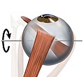
temporal side of the eyeball. This insertion is around 7 mm from the corneal limbus. It has a width of around 10 mm. The lateral rectus is the only muscle...
7 KB (751 words) - 17:22, 4 May 2024
several ways: Distortion of the corneal optics. This begins usually when the pterygium is greater than 2mm from the corneal limbus. Disruption of the tear. The...
6 KB (691 words) - 15:19, 5 March 2024

orbit (left/right) annulus of Zinn at orbital apex 7.5 mm superior to corneal limbus ophthalmic artery oculomotor nerve [CNIII], superior branch adducts...
138 KB (921 words) - 03:49, 31 July 2024

opposed to the near-uniform thickness and parallel arrangement of the corneal collagen. Moreover, the cornea bears more mucopolysaccharide (a carbohydrate...
16 KB (2,032 words) - 05:55, 25 July 2024

thin membrane which envelops the eyeball from the optic nerve to the corneal limbus, separating it from the orbital fat and forming a socket in which it...
5 KB (598 words) - 13:58, 11 September 2023
Descemet membrane endothelial keratoplasty (category Corneal transplantation)
incisions just anterior to the corneal limbus. The donor tissue is tamponaded against the person's exposed posterior corneal stroma by injecting a small...
3 KB (252 words) - 02:27, 31 October 2023
This could result in the early development of arcus lipoides, hazy corneal limbus, and hyperopia. There is evidence that cornea plana 2 is caused by mutations...
8 KB (717 words) - 10:54, 7 March 2024

insertion has a width of around 10.5 mm. It is around 7 mm from the corneal limbus. The inferior rectus muscle is supplied by an inferior muscular branch...
7 KB (688 words) - 14:29, 3 May 2024

performed by Custer et al.: The conjunctival peritomy is performed at the corneal limbus, preserving as much healthy tissue as possible. Anterior Tenon's fascia...
41 KB (4,944 words) - 12:14, 13 August 2024
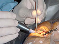
incision into the anterior chamber of the eye is made at or near the corneal limbus, where the cornea and sclera meet, either superior or temporal. Advantages...
150 KB (15,139 words) - 21:21, 18 August 2024

in the cartilage of the ear and other cartilage, and the sclera and corneal limbus of the eye. After the age of 30, people begin to develop pain in the...
19 KB (2,158 words) - 01:25, 18 June 2024

iris resulting from a manifestation of the optical properties of the corneal limbus. Limbal rings are not present in all individuals, and their thickness...
66 KB (7,576 words) - 03:05, 29 August 2024

conjunctiva is the thin transparent tissue that covers the eye from the Corneal limbus to the lid margin. Many conditions can lead to the inflammation of the...
5 KB (621 words) - 10:37, 27 October 2023

five-sixths; its diameter is typically about 24 mm (0.94 in). An area termed the limbus connects the cornea and sclera. The iris is the pigmented circular structure...
77 KB (9,246 words) - 23:26, 24 August 2024
incision into the anterior chamber of the eye is made at or near the corneal limbus, where the cornea and sclera meet, either superior or temporal. Advantages...
43 KB (4,789 words) - 00:53, 9 May 2024
This insertion has a width of around 11 mm. It is around 8 mm from the corneal limbus. The superior rectus muscle is supplied by the superior division of...
9 KB (942 words) - 14:27, 3 May 2024

brown, golden, or reddish-green, are 1 to 3 mm wide, and appear at the corneal limbus. They do not occur in all people with Wilson's disease and may be seen...
42 KB (4,960 words) - 11:07, 13 August 2024
the limbus at a 60°angle. The anterior chamber drainage angle is then graded as a ratio between the peripheral anterior chamber depth and corneal thickness...
11 KB (1,311 words) - 08:52, 5 June 2024

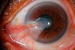
Corneal neovascularization (CNV) is the in-growth of new blood vessels from the pericorneal plexus into avascular corneal tissue as a result of oxygen...
14 KB (1,530 words) - 14:10, 19 June 2024

thickened accumulation of tissue around limbus and presence of discrete whitish raised dots along the limbus (Tranta's spots). Mixed form- Shows the features...
7 KB (709 words) - 13:57, 14 February 2024
Corneosclera with 8. Cornea, 9. Trabecular meshwork and Schlemm's canal. 10. Corneal limbus and 11. Sclera; 12. Conjunctiva, 13. Uvea with 14. Iris, 15. Ciliary...
29 KB (3,531 words) - 02:34, 18 August 2024