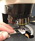
Confocal microscopy, most frequently confocal laser scanning microscopy (CLSM) or laser scanning confocal microscopy (LSCM), is an optical imaging technique...
46 KB (5,298 words) - 15:29, 17 September 2024
light microscopy and transmission electron microscopy) or by scanning a fine beam over the sample (for example confocal laser scanning microscopy and scanning...
69 KB (8,315 words) - 10:01, 9 July 2024
results in the spatial resolution of the image. This contrasts with confocal microscopy, where the spatial resolution is produced by the interaction of excitation...
34 KB (3,475 words) - 19:51, 14 August 2024
to about a factor of two) beyond the diffraction-limit, such as confocal microscopy with closed pinhole or aided by computational methods such as deconvolution...
88 KB (10,141 words) - 17:32, 4 September 2024
comparable to confocal microscopy. Because light sheet fluorescence microscopy scans samples by using a plane of light instead of a point (as in confocal microscopy)...
41 KB (4,599 words) - 15:54, 17 September 2024
Tomography (redirect from Synchroton-radiation X-ray tomographic microscopy)
W. Z. W. (December 2015). "Three dimensions localization of tumors in confocal microwave imaging for breast cancer detection" (PDF). Microwave and Optical...
19 KB (1,693 words) - 10:20, 1 July 2024
Scanning confocal electron microscopy (SCEM) is an electron microscopy technique analogous to scanning confocal optical microscopy (SCOM). In this technique...
7 KB (709 words) - 14:25, 16 September 2023
Fluorescence microscope (redirect from Flourescence microscopy)
more complex fluorescence microscopy techniques like confocal microscopy and total internal reflection fluorescence microscopy while xenon lamps, and mercury...
25 KB (2,745 words) - 10:17, 9 August 2024
lens is the same as one focus of the next lens. Confocal laser scanning microscopy Confocal microscopy List of orbits § Eccentricity classifications This...
991 bytes (158 words) - 20:47, 17 December 2020
fluorescence, it can achieve resolution better than traditional confocal microscopy. Normal fluorescence occurs by exciting an electron from the ground...
33 KB (3,866 words) - 17:27, 8 June 2024
4Pi microscope (redirect from 4Pi microscopy)
spherical focal spot with 5–7 times less volume than that of standard confocal microscopy. The improvement in resolution is achieved by using two opposing...
9 KB (1,149 words) - 09:41, 14 July 2024
samples – confocal microscopy and widefield microscopy – both have significant drawbacks for this type of application. In widefield microscopy, both in-focus...
21 KB (2,784 words) - 03:19, 26 August 2024
theory of evolution) and new technologies have become available (confocal microscopy, DNA sequencing, genomics). Additionally, the discovery of Onychophora...
10 KB (1,014 words) - 13:23, 6 November 2023
tissue which otherwise is only possible by tissue biopsy. Similar to confocal microscopy, the laser in CLE filtered by the pinhole excites the fluorescent...
28 KB (2,824 words) - 01:37, 24 June 2024
multiphoton microscopy is required. Multiphoton microscopy provides considerably greater depth of penetration than single-photon confocal microscopy. Multiphoton...
13 KB (1,411 words) - 00:48, 15 May 2024
Histology (section Light microscopy)
Fluorescence microscopy and confocal microscopy are used to detect fluorescent signals with good intracellular detail. For electron microscopy heavy metals...
33 KB (3,197 words) - 22:58, 11 September 2024
sample. It can be used as an imaging technique in confocal microscopy, two-photon excitation microscopy, and multiphoton tomography. The fluorescence lifetime...
24 KB (3,092 words) - 21:09, 11 July 2024
photoacoustic/thermoacoustic tomography, i.e., PAT/TAT) and photoacoustic microscopy (PAM), have been developed. A typical PAT system uses an unfocused ultrasound...
14 KB (1,715 words) - 07:59, 6 May 2024
to point scanning techniques such as laser scanning confocal microscopy) fluorescence microscopy imaging methods that allow obtaining images with a resolution...
27 KB (3,213 words) - 22:07, 18 May 2024
Endomicroscopy (category Microscopy)
‘optical biopsy’. It generally refers to fluorescence confocal microscopy, although multi-photon microscopy and optical coherence tomography have also been...
12 KB (1,703 words) - 01:47, 11 May 2024
Optical microscope (redirect from Optical microscopy)
analysis of surface structures) Fluorescence microscopy, both: Epifluorescence microscopy Confocal microscopy Microspectroscopy (where a UV-visible spectrophotometer...
52 KB (5,988 words) - 22:59, 21 August 2024
Raman microscope (redirect from Raman microscopy)
numerical aperture of the focusing element. In confocal Raman microscopy, the diameter of the confocal aperture is an additional factor. As a rule of...
17 KB (1,912 words) - 19:00, 20 September 2023
Neuromorphology (section Confocal microscopy)
of techniques have been used to study neuromorphology, including confocal microscopy, design-based stereology, neuron tracing and neuron reconstruction...
17 KB (2,037 words) - 23:36, 23 April 2024
us try to understand the principle of confocal microscopy in terms of momentum basis, here. In confocal microscopy, the effect of the pinhole can be understood...
6 KB (1,154 words) - 02:52, 30 January 2023
Live-cell imaging (category Microscopy)
103828. Keller HE (2006), "Objective Lenses for Confocal Microscopy", Handbook of Biological Confocal Microscopy, Springer US, pp. 145–161, doi:10.1007/978-0-387-45524-2_7...
30 KB (3,419 words) - 12:08, 9 July 2024
for large enough (micrometer-sized) particles via traditional or confocal microscopy. The radial distribution function is of fundamental importance since...
30 KB (4,541 words) - 15:36, 21 September 2024
detection methods include photography, dermatoscopy, sonography, confocal microscopy, Raman spectroscopy, fluorescence spectroscopy, terahertz spectroscopy...
58 KB (6,460 words) - 16:39, 24 September 2024
the 1970s, but was later shown by new techniques developed for electron microscopy to be simply an artifact of chemical fixation. Standardization of fixation...
19 KB (2,387 words) - 06:19, 26 November 2023
interferometry, digital holography, confocal microscopy, focus variation, structured light, electrical capacitance, electron microscopy, photogrammetry and non-contact...
19 KB (2,232 words) - 07:22, 19 May 2024
Three-dimensional projection of a mammalian Golgi stack imaged by confocal microscopy and volume surface rendered using Imaris software. Taken from the...
25 KB (2,765 words) - 02:05, 30 September 2024


