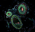
Immunofluorescence (IF) is a light microscopy-based technique that allows detection and localization of a wide variety of target biomolecules within a...
21 KB (2,054 words) - 16:47, 21 April 2024
quantifying ANAs are indirect immunofluorescence and enzyme-linked immunosorbent assay (ELISA). In immunofluorescence, the level of autoantibodies is...
54 KB (6,097 words) - 21:43, 28 June 2024
Direct fluorescent antibody (redirect from Direct immunofluorescence)
A direct fluorescent antibody (DFA or dFA), also known as "direct immunofluorescence", is an antibody that has been tagged in a direct fluorescent antibody...
3 KB (410 words) - 12:55, 16 December 2020
diagnosed with the aid of immunofluorescence studies. Cutaneous conditions with positive direct or indirect immunofluorescence when using salt-split skin...
2 KB (95 words) - 17:03, 14 March 2022
Anti-dsDNA antibodies (section Immunofluorescence)
Blood tests such as enzyme-linked immunosorbent assay (ELISA) and immunofluorescence are routinely performed to detect anti-dsDNA antibodies in diagnostic...
23 KB (2,924 words) - 07:24, 20 July 2024
Pemphigoid (section Direct immunofluorescence)
histopathology and direct immunofluorescence, indirect immunofluorescence and the ELISA test. Among all, direct immunofluorescence is the gold standard for...
50 KB (5,853 words) - 12:52, 2 November 2024
specialized testing needs to be performed on biopsies, including immunofluorescence, immunohistochemistry, electron microscopy, flow cytometry, and molecular-pathologic...
51 KB (5,579 words) - 13:13, 10 November 2024
cerebellum with bound anti-GAD65 monoclonal antibodies (green), indirect immunofluorescence Specialty Neurology Symptoms muscular rigidity and trigger-induced...
27 KB (2,949 words) - 01:40, 29 October 2024
Immunofluorescence micrograph of three cytotoxic T cells (outer three) surrounding a cancer cell. Lytic granules (red) are secreted at the contact site...
14 KB (1,377 words) - 16:49, 20 November 2024
Immunofluorescence pattern of SS-A and SS-B antibodies. Produced using serum from a patient on HEp-20-10 cells with a FITC conjugate....
14 KB (1,783 words) - 14:40, 11 November 2024
non-organ- and non-species-specific anti-organelle antibody detected by immunofluorescence: the mitochondrial antibody number 5. Clin Exp Immunol 31(3):357-366...
5 KB (500 words) - 10:03, 21 October 2023
basement membrane without a hyperproliferation of the glomerular cells. Immunofluorescence demonstrates diffuse granular uptake of IgG. The basement membrane...
21 KB (2,134 words) - 13:51, 22 September 2024
proliferation or crescentic glomerulonephritis may also be present. Immunofluorescence shows mesangial deposition of IgA often with C3 and properdin and...
29 KB (3,482 words) - 09:34, 27 July 2024
Immunofluorescence image of HeLa cells grown in tissue culture and stained with antibody to actin in green, vimentin in red and DNA in blue...
49 KB (5,685 words) - 15:26, 23 October 2024
immunohistochemistry, or when the stain is a fluorescent molecule, immunofluorescence. This technique has greatly increased the ability to identify categories...
33 KB (3,193 words) - 04:44, 16 November 2024
assistant divides the specimen(s) for submission for light microscopy, immunofluorescence microscopy and electron microscopy. The pathologist will examine the...
62 KB (6,985 words) - 10:25, 17 November 2024
crescents will be normally observed. When these are subjected to immunofluorescence, three patterns can be observed: linear, granular and negative (pauci-immune)...
3 KB (291 words) - 04:33, 2 February 2024
severity of portal hypertension. Main antinuclear antibody patterns on immunofluorescence A schematic representation of an antibody JB Imboden, DB Hellmann...
3 KB (269 words) - 00:59, 5 February 2024
direct immunofluorescence microscopy (DIF) on a skin biopsy specimen, and (3) positive epidermal side staining by indirect immunofluorescence microscopy...
13 KB (1,333 words) - 22:26, 8 October 2024
become the primary diagnostic tool in the future.[citation needed] Immunofluorescence or immunoperoxidase assays are commonly used to detect whether a virus...
11 KB (1,346 words) - 12:41, 4 January 2024
Main antinuclear antibody patterns on immunofluorescence. CREST syndrome typically displays the centrome pattern....
8 KB (771 words) - 21:24, 12 August 2024
Fluorescence microscope (section Immunofluorescence)
different molecule which binds the target of interest within the sample. Immunofluorescence is a technique which uses the highly specific binding of an antibody...
25 KB (2,729 words) - 08:21, 10 November 2024
Inside the cell, the cadherins are linked to actin filaments. In immunofluorescence microscopy, the actin filament network appears as a thick border surrounding...
28 KB (3,056 words) - 06:49, 2 November 2024
Berliner, Norman Jones and Hugh J Creech was the first to develop immunofluorescence in 1941. This led to the later development of immunohistochemistry...
33 KB (3,561 words) - 16:36, 24 November 2024
done via antibody staining, hemadsorption using red blood cells, or immunofluorescence microscopy. Shell vial cultures, which can identify infection via...
114 KB (13,208 words) - 20:42, 2 November 2024
Lupus band test is done upon skin biopsy, with direct immunofluorescence staining, in which, if positive, IgG and complement depositions are found at the...
3 KB (202 words) - 17:41, 13 January 2022
antigen-antibody are used to diagnose infectious diseases, for example ELISA, immunofluorescence, Western blot, immunodiffusion, immunoelectrophoresis, and magnetic...
121 KB (13,516 words) - 08:01, 2 November 2024
and the optical microscope. Developments in electron microscopy, immunofluorescence, and the use of frozen tissue-sections have enhanced the detail that...
24 KB (3,035 words) - 16:00, 19 November 2024
Three-Dimensional Imaging with Confocal Laser Scanning Microscopy and Double Immunofluorescence". Modern Pathology. 14 (9): 854–861. doi:10.1038/modpathol.3880401...
39 KB (3,021 words) - 01:53, 19 November 2024
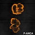
systemic vasculitis, so called ANCA-associated vasculitides (AAV). Immunofluorescence (IF) on ethanol-fixed neutrophils is used to detect ANCA, although...
20 KB (2,272 words) - 06:48, 24 August 2024

