Confocal microscopy, most frequently confocal laser scanning microscopy (CLSM) or laser scanning confocal microscopy (LSCM), is an optical imaging technique...
46 KB (5,298 words) - 06:21, 9 April 2024
In geometry, confocal means having the same foci: confocal conic sections. For an optical cavity consisting of two mirrors, confocal means that they share...
991 bytes (156 words) - 20:47, 17 December 2020
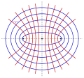
called confocal if they have the same foci. Because ellipses and hyperbolas have two foci, there are confocal ellipses, confocal hyperbolas and confocal mixtures...
22 KB (3,676 words) - 13:31, 11 January 2024

Confocal endoscopy, or confocal laser endomicroscopy (CLE), is a modern imaging technique that allows the examination of real-time microscopic and histological...
28 KB (2,824 words) - 01:37, 24 June 2024
Scanning confocal electron microscopy (SCEM) is an electron microscopy technique analogous to scanning confocal optical microscopy (SCOM). In this technique...
7 KB (709 words) - 14:25, 16 September 2023

Rayleigh length (redirect from Confocal parameter)
where the area of the cross section is doubled. A related parameter is the confocal parameter, b, which is twice the Rayleigh length. The Rayleigh length is...
4 KB (492 words) - 16:00, 7 February 2024

Ellipsoidal coordinates (redirect from Confocal ellipsoidal coordinates)
quadratic coordinate surfaces, the ellipsoidal coordinate system is based on confocal quadrics. The Cartesian coordinates ( x , y , z ) {\displaystyle (x,y,z)}...
7 KB (1,149 words) - 01:54, 3 June 2024

head-mounted graphical display (1963) and the confocal microscope (1957, a predecessor to today's widely used confocal laser scanning microscope). He developed...
34 KB (3,010 words) - 23:17, 22 July 2024

like an epifluorescence microscope or a more complicated design such as a confocal microscope, which uses optical sectioning to get better resolution of the...
25 KB (2,745 words) - 10:17, 9 August 2024

microscopy and other imaging techniques such as 3D microscopy (like in confocal laser scanning microscopy) and fluorescence microscopy. The degree of spreading...
24 KB (3,265 words) - 14:04, 25 June 2024


Microscopy (section Confocal)
electron microscopy) or by scanning a fine beam over the sample (for example confocal laser scanning microscopy and scanning electron microscopy). Scanning probe...
69 KB (8,315 words) - 10:01, 9 July 2024

which results in the spatial resolution of the image. This contrasts with confocal microscopy, where the spatial resolution is produced by the interaction...
34 KB (3,475 words) - 19:51, 14 August 2024
Paraboloidal coordinates (redirect from Confocal paraboloidal coordinates)
3rd print ed.). New York: Springer-Verlag. pp. 44–48 (Table 1.11). ISBN 978-0-387-18430-2. MathWorld description of confocal paraboloidal coordinates...
9 KB (1,524 words) - 19:19, 28 April 2024

deactivates the fluorescence, it can achieve resolution better than traditional confocal microscopy. Normal fluorescence occurs by exciting an electron from the...
33 KB (3,866 words) - 17:27, 8 June 2024
Animated confocal micrograph of part of a biological neural network in a mouse's striatum...
7 KB (759 words) - 12:22, 28 July 2024
Focus (geometry) (section Confocal curves)
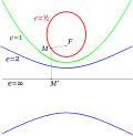
In geometry, focuses or foci (/ˈfoʊkaɪ/; sg.: focus) are special points with reference to which any of a variety of curves is constructed. For example...
10 KB (1,424 words) - 22:19, 18 October 2023

applicable to specific systems, e.g., Xe bubbles in aluminum. Scanning confocal electron energy loss microscopy (SCEELM) is a new analytical microscopy...
19 KB (2,262 words) - 17:09, 17 May 2024
ISO 25178 (section Chromatic confocal gauge)
(stylus) instruments Part 602: Nominal characteristics of non-contact (confocal chromatic probe) instruments Part 603: Nominal characteristics of non-contact...
11 KB (1,293 words) - 18:48, 12 March 2024

Mouse brain tissue, fixed via perfusion, stained via immunohistochemistry and imaged using confocal microscopy....
19 KB (2,387 words) - 06:19, 26 November 2023

radius equal to the cavity length. A common and important design is the confocal resonator, with mirrors of equal radii to the cavity length (R1 = R2 =...
18 KB (2,326 words) - 09:26, 5 July 2024

two-dimensional orthogonal coordinate system in which the coordinate lines are confocal parabolas. A three-dimensional version of parabolic coordinates is obtained...
8 KB (1,109 words) - 11:44, 7 June 2024
modestly (up to about a factor of two) beyond the diffraction-limit, such as confocal microscopy with closed pinhole or aided by computational methods such as...
87 KB (10,123 words) - 03:18, 29 July 2024
Neuromorphology (section Confocal microscopy)
variety of techniques have been used to study neuromorphology, including confocal microscopy, design-based stereology, neuron tracing and neuron reconstruction...
17 KB (2,037 words) - 23:36, 23 April 2024
Animated confocal micrograph, showing interconnections of medium spiny neurons in mouse striatum...
12 KB (1,342 words) - 21:06, 3 June 2024

microscopy drove the development of a major modern microscope design, the confocal microscope. The principle was patented in 1957 by Marvin Minsky, although...
31 KB (3,699 words) - 15:02, 20 May 2024
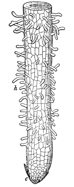
Calcofluor White confocal fluorescent image of developing tomato root...
10 KB (1,207 words) - 20:20, 27 June 2024

and Vimentin in Malignant Rhabdoid Tumor: Three-Dimensional Imaging with Confocal Laser Scanning Microscopy and Double Immunofluorescence". Modern Pathology...
39 KB (3,016 words) - 15:08, 13 July 2024

W. Z. W. (December 2015). "Three dimensions localization of tumors in confocal microwave imaging for breast cancer detection" (PDF). Microwave and Optical...
19 KB (1,693 words) - 10:20, 1 July 2024