ملف:Hippel Lindau.gif - ويكيبيديا
الملف الأصلي (805 × 949 بكسل حجم الملف: 278 كيلوبايت، نوع MIME: image/gif)

ملخص

الوصف
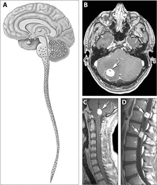
English: Distribution of Hemangioblastomas in the Central Nervous Systems of Study Patients

(A) Schematic representation of the distribution of CNS hemangioblastomas (red dots) in the 25 von Hippel-Lindau disease patients on MRI. Most (98%) of hemangioblastomas were found below the level of the tentorium in the cerebellum, brainstem, and spinal cord.

(B–D) Contrast-enhanced MRI demonstrating representative locations of hemangioblastomas including the cerebellum (B), brainstem (C) and spinal cord (D). (B) Axial view through the cerebellum demonstrating a hyperintense enhancing hemangioblastoma (arrow) with surrounding edema (hypointense area surrounding the tumor) that frequently is associated with these lesions. (C) Sagittal view through the posterior fossa demonstrating a hyperintense enhancing brainstem (medullary) hemangioblastoma (arrow) with surrounding edema. (D) Sagittal view through the thoracic and lumbar spinal cord demonstrating two hyperintense enhancing hemangioblastomas (arrows). The superior tumor is associated with a large intraspinal cyst (syrinx) that is common with these neoplasms (arrowhead)
المصدر http://medicine.plosjournals.org/perlserv/?request=get-document&doi=10.1371/journal.pmed.0040060
المؤلف

ترخيص

w:ar:مشاع إبداعي
نسب العمل إلى مُؤَلِّفه
يحقُّ لك:
  • مشاركة العمل – نسخ العمل وتوزيعه وبثُّه
  • إعادة إنتاج العمل – تعديل العمل
حسب الشروط التالية:
  • نسب العمل إلى مُؤَلِّفه – يلزم نسب العمل إلى مُؤَلِّفه بشكل مناسب وتوفير رابط للرخصة وتحديد ما إذا أجريت تغييرات. بالإمكان القيام بذلك بأية طريقة معقولة، ولكن ليس بأية طريقة تشير إلى أن المرخِّص يوافقك على الاستعمال.
PLOS
PLOS
نُشِر هذا الملفُّ في دورية المكتبة العامَّة للعلوم. يُصرِّح الموقع الرسميِّ بأنَّ مُحتويات دوريات المكتبة العامَّة للعلوم مَنشورةٌ جميعها تحت رخصة المَشاع الإِبداعيِّ نسبة المُصنَّف إِلى مُؤَلِّفه (أو إلى إصداراتٍ سابِقة مِن الرُّخصة حسب تاريخ الإِصدار)، ما لم يذكر خلاف ذلك.

الشروحات

أضف شرحاً من سطر واحد لما يُمثِّله هذا الملف

العناصر المصورة في هذا الملف

يُصوِّر

تاريخ الملف

اضغط على زمن/تاريخ لرؤية الملف كما بدا في هذا الزمن.

زمن/تاريخصورة مصغرةالأبعادمستخدمتعليق
حالي13:43، 31 مايو 2007تصغير للنسخة بتاريخ 13:43، 31 مايو 2007805 × 949 (278 كيلوبايت)Filip emDistribution of Hemangioblastomas in the Central Nervous Systems of Study Patients (A) Schematic representation of the distribution of CNS hemangioblastomas (red dots) in the 25 von Hippel-Lindau disease patients on MRI. Most (98%) of hemangioblastomas w

الصفحة التالية تستخدم هذا الملف:

الاستخدام العالمي للملف

الويكيات الأخرى التالية تستخدم هذا الملف: