File:FLAIR MRI of meningitis.jpg

FLAIR_MRI_of_meningitis.jpg (600 × 521 pixels, file size: 131 KB, MIME type: image/jpeg)

Summary

Description
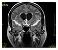
English: Postcontrast Fluid-attenuated inversion recovery (FLAIR) MRI of a case of meningitis. It shows the enhancement of meninges at the tentorium and in the parietal region, with evidence of dilated ventricles.
Date
Source (2014). "Diagnostic Accuracy of Contrast-Enhanced FLAIR Magnetic Resonance Imaging in Diagnosis of Meningitis Correlated with CSF Analysis". ISRN Radiology 2014: 1–7. DOI:10.1155/2014/578986. ISSN 2314-4084. (CC-BY-3.0)
Author Aneel Kumar Vaswani, Waseem Mehmood Nizamani, Muhammad Ali, Geeta Aneel, Bhesham Kumar Shahani, and Sajjad Hussain
Uploaded by Mikael Häggström

Licensing

w:en:Creative Commons
attribution
This file is licensed under the Creative Commons Attribution 3.0 Unported license.
You are free:
  • to share – to copy, distribute and transmit the work
  • to remix – to adapt the work
Under the following conditions:
  • attribution – You must give appropriate credit, provide a link to the license, and indicate if changes were made. You may do so in any reasonable manner, but not in any way that suggests the licensor endorses you or your use.

Captions

Add a one-line explanation of what this file represents

Items portrayed in this file

depicts

20 March 2014

File history

Click on a date/time to view the file as it appeared at that time.

Date/TimeThumbnailDimensionsUserComment
current13:47, 15 October 2017Thumbnail for version as of 13:47, 15 October 2017600 × 521 (131 KB)Mikael Häggström{{Information |Description ={{en|1=Postcontrast Fluid-attenuated inversion recovery (FLAIR) MRI of a case of meningitis. It shows the enhancement of meninges at the tentorium and in the...

Global file usage