File:Sucking chest wound mechanics 2.jpg

Sucking_chest_wound_mechanics_2.jpg (248 × 419 pixels, file size: 34 KB, MIME type: image/jpeg)

Description
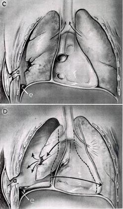
English: caption reads: FIGURE 2.—Continued. C. Packing of sucking wound (a), after which respiration becomes more normal. Hemothorax (e). D. Development of tension pneumothorax because air cannot escape from tear in lung (a), after wound is adequately packed. If it develops, it must be treated by closed (catheter) drainage of cavity. Hemothorax (e).
Date Date of drawing is unknown, year of publication is 1965.
Source Surgery in World War II: Thoracic surgery, Volume II (1965). Medical department, United States Army.CHAPTER I: Special Types of Thoracic Wounds. Direct link: [1]
Author Lyman A. Brewer III, M.D., and Thomas H. Burford, M.D.
Permission
(Reusing this file)
pd - US Army
Other versions Image:Sucking chest wound mechanics.jpg
Public domain
This file is a work of a U.S. Army soldier or employee, taken or made as part of that person's official duties. As a work of the U.S. federal government, it is in the public domain in the United States.

العربية  বাংলা  català  čeština  Deutsch  English  español  eesti  فارسی  suomi  français  hrvatski  magyar  Bahasa Indonesia  italiano  日本語  한국어  lietuvių  македонски  മലയാളം  မြန်မာဘာသာ  Nederlands  polski  português  русский  sicilianu  српски / srpski  Türkçe  українська  Tiếng Việt  中文(简体)  中文(繁體)  +/−

Captions

Add a one-line explanation of what this file represents

Items portrayed in this file

depicts

File history

Click on a date/time to view the file as it appeared at that time.

Date/TimeThumbnailDimensionsUserComment
current06:10, 14 June 2008Thumbnail for version as of 06:10, 14 June 2008248 × 419 (34 KB)Delldot{{Information |Description={{en|1=caption reads: FIGURE 2.—Continued. C. Packing of sucking wound (a), after which respiration becomes more normal. Hemothorax (e). D. Development of tension pneumothorax because air cannot escape from tear in lung (a), a

The following page uses this file:

Metadata